At a time when technology is reshaping how we diagnose, a patient with an autoimmune disorder compels a doctor to turn to old school hacks

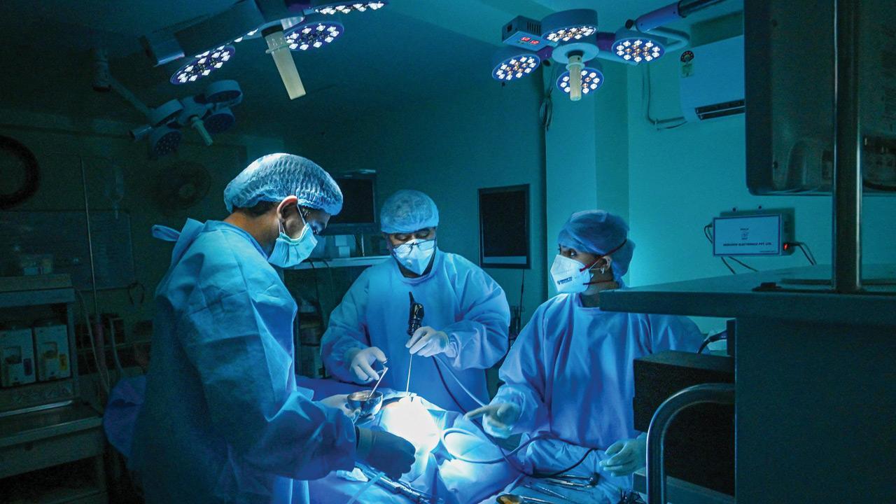
Representation pic
 Robert came to us from southern Africa. He had had surgery on his cervical spine one year ago for a compression of his spinal cord that had made it difficult for him to use his hands and legs. He made an exemplary recovery from his symptoms and regained his lost function in a span of eight months.
ADVERTISEMENT
However, he began to notice something funny. His voice began to undergo a consistent change; it started developing a nasal twang, the kind that would make one believe he was from the Philippines.
Within a few weeks, he also found it arduous to swallow food and even drink water. He choked on tiny morsels of grain; food would sometimes make its way down the windpipe instead of the food pipe, which would cause him to asphyxiate. The doctors back home inserted a nasogastric tube prohibiting him from consuming anything orally. They even made a hole in his wind pipe, doing a tracheostomy to safeguard him from suffocating in his own secretions. He lived this way for an entire year before they sent him to us to figure out what was wrong. It was puzzling; an MRI of the brain was clear and the spine too had been adequately decompressed at the level they operated on, but there was some mild stenosis above. They told him he might need more surgery and put him on a plane.
Also Read: The surgical football
When I first saw him, he looked emaciated, with a tube stuck in his nose and another one popping out of his throat, which he blocked with a finger for his voice to be audible. He was six-foot-tall and weighed about 50 kg; he had lost half his weight in the past few months. “Why does one of your eyes look a little smaller than the other?” I asked after listening to his entire ordeal. His wife jumped off the couch to check on my observation, and after peering closely, agreed. “The left one, correct?” she asked, surprised at how she hadn’t noticed it in the past year. “Does that mean anything?” she continued, perturbed. I explained that there is always some subtle asymmetry in the human body: often, one breast or testicle is slightly smaller than the other, or one half of the brain is slightly larger than the other, and then there are people who have an indistinct difference in the size of their hands or feet, and sometimes even the eyes. The key is in understanding what is clinically significant to our diagnosis.
“Do any of your symptoms worsen as your day progresses?” I enquired further. “Yes,” he said emphatically, once again blocking his tube to be heard, the voice becoming more nasal, the fatigue more pronounced. I told them I had an idea of what we were dealing with and that I needed to check with our neurologist, who confirmed the diagnosis in a few minutes of seeing him. “Without a doubt, this is myasthenia gravis,” he said with an air of authority, explaining to Robert that the disorder indicated grave or serious muscle weakness. “This is a disease in which antibodies are made by a person’s immune system to prevent certain nerve muscle interactions,” he continued.
“This affects certain groups of muscles causing them to get weak, more so as the day progresses.” Turning to me, he announced, “We’ll give him a few drugs to block the antibodies and he’ll be okay in no time!” his excitement palpable as he snapped his fingers at me.
There is an exhilaration in recognising what is staring you in the face. The joy of making a concrete clinical diagnosis in medicine in today’s AI-driven metaverse is a simple example of that. We now live in a time where images and investigations show us the way forward, but once in a while a Robert comes along to keep the light aflame; one clear look at his eye told us what was going on.
Senior clinicians look back lamentingly on the days when they spent hours getting to know a patient, sitting at their bedside and talking to them gently, examining them with excruciating detail, until they were able to discern what was going on, only to be thrilled at making the correct diagnosis and seeing the results of their treatment pan out in front of them. Today, most of us glance at reports and films to know how to move forward and focus on technical nuances. After all, it is also imperative that we use all the modern technology available to us to enhance our surgical outcomes.
The eye is a direct extension of the brain. Looking into someone’s eyes gives us so much information—medically, emotionally, and spiritually. You can diagnose something as simple as a cataract, conjunctivitis or jaundice, copper deposition (indicating liver disease) or thyroid dysfunction, or something more sinister such as a retinal detachment or neuritis. Looking into the eye can tell you if there is raised pressure inside the brain, and if you look a little deeper, you can even diagnose increased pressure inside the heart. The important thing is to look hard enough. Looking into patients’ eyes while talking to them builds trust and confidence. It allows them to share their real story. It tells us parts of their story that words fail to express.
The eye is also a surgical corridor to the brain. Instead of sawing through the skull bone, for certain pathologies, we can make a tiny incision behind or through the eyelid and then make a small hole through the eye socket to reach the brain. We like to call it ‘natural orifice’ surgery. I’m not sure if this makes the idea of brain surgery less or more terrifying.
Robert’s diagnosis was confirmed with a few lab tests and nerve conduction studies. He was started on a cocktail of medication by the neurologist that improved his voice and strengthened the muscles he needed to eat, chew, and swallow within a matter of days. His eye opened up like it was ready to bloom. We removed his nasogastric tube and closed his tracheostomy over the next two weeks. His smile had never been broader because he was able to eat like a normal human being for the first time in a year; he even polished off the food that came for his wife. He put on 9 kg while he was admitted with us.
It was the perfect start to the new year.
The writer is practicing neurosurgeon at Wockhardt Hospitals and Honorary Assistant Professor of Neurosurgery at Grant Medical College and Sir JJ Group of Hospitals.
 Subscribe today by clicking the link and stay updated with the latest news!" Click here!


